


Medicine and Microscopes: How Microscopes Have Impacted the Healthcare Field
Microscopes are fantastically versatile tools. When we are children, they open our eyes to secret worlds within a drop of water or reveal the alien-looking structure of a fly’s eye. In university settings, they help students identify the parts of a cell or examine tissue samples. Hobbyists and researchers alike can take handheld digital microscopes out into the field to conveniently analyze plant, animal or mineral matter on the go. Microscopes cast a wide net in their usefulness, but one of the most important ways in which they touch our lives is in medicine. In this article, we explore how microscopes have greatly impacted the healthcare field through the ages.
The First Microscopes
In the 17th century, Antoine van Leeuwenhoek improved upon primitive versions of the microscope by placing a small spherical glass lens in an adjustable shaft that could be manipulated to focus on minute specimens. He was able to see red blood cells and microorganisms, which led to him being recognized as the “father of microbiology.” While microscopes certainly had a long way to go from Leeuwenhoek’s device to what we use today, their importance in healthcare has been evident almost since their inception.
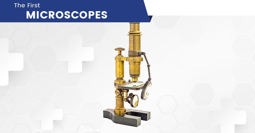
Leeuwenhoek and other early microscopists recorded bacteria, protozoa, epithelial cells and other then-mysteries of the human body, entering their discoveries into medical reference books. Still, it would be another hundred years before the importance of microscopes in medicine was universally acknowledged and they would become indispensable both in clinical settings and at universities around the world.
Medical Education
When you think of a microscope, one of the first images that comes to mind may be a scientist in a white lab coat, bending over an eyepiece. This isn’t too far from the truth. Healthcare education relies heavily on the use of medical microscopes to teach students how to identify a myriad of maladies that can be found in blood or tissue samples. From your first basic biology class you take at your university, all the way up through your pre-med course load, if you embark on the educational path to be a healthcare provider, you will encounter microscopes along the way.
Teaching students how to use microscopes at school can help them identify microbes causing infections once they enter the medical field. But learning how to use the microscope is only one side of the coin. Educators can utilize digital microscopes with cameras to project images for the entire classroom to see. Digital microscopes use optics and a digital camera to relay images onto a computer. Still images and videos can be projected in real-time so an entire lecture hall can observe the same specimen. Teachers use these devices to ensure everyone views the exact same image, avoiding the variations that arise from students taking turns to look into an eyepiece individually. This use of USB technology–where images can be saved and shared between users–enables students to reference the data later as they continue their studies.

Clinical Settings
Before microscopes found their foothold in modern medicine, people–even medical doctors–believed that God or evil spirits sent diseases to punish humans. In the 1860s, Louis Pasteur developed a theory that “germs” were actually the culprit behind our illnesses. Pasteur noticed correlations between specific diseases and the objects he observed in sick patients’ blood. We now know that the “germs” Pasteur referred to were really bacteria, viruses and other pathogens. His observations paved the way for modern medicine to identify, treat and even prevent infectious diseases.
Bacteria and blood cells come to light in phase microscopes, which are used in many clinical settings. If you took a biology class in high school, you may remember swabbing the inside of your cheek with a flat toothpick and placing the tissue on a slide. The cells that appeared under the microscope were epithelial cells, which line the surfaces of our organs and blood vessels. Doctors and clinicians still use medical microscopes to identify these types of cells, which can often tell us when something is going wrong in our bodies.
Types of Microscopes in Clinical Settings
There are many reasons why healthcare providers might need to use a microscope. Also, it can take more than just one type of microscope to accommodate each of these functions, so hospitals and doctors’ offices often have a wide variety of types of scopes on hand. One of the most common microscopes is the light microscope. These use light to illuminate an image, while one or sometimes multiple lenses magnify the specimen. These are used to examine tissue or identify unicellular organisms, like those that can be found in our blood and indicate illness.
Fluorescent microscopes use ultraviolet light to illuminate greater cellular detail. By blocking outside light, these microscopes can increase the resolution of an image and show the specimen in three dimensions. These devices are excellent for identifying bacteria in a sample. Electron microscopes use concentrated beams of electrons and dyes to illuminate their specimens. These electron beams have very short wavelengths, which means they are able to produce exceptionally high-resolution images. Clinics can utilize these scopes to identify and examine viruses within the bloodstream.

Somewhere in between light and electron microscopes lie X-ray microscopes. X-rays are very compact beams that cannot be detected by the human eye. They also do not reflect or refract easily, which means they can produce very clear, detailed images. They have an extremely high resolution–enough to allow the observer to hone in on individual atoms–and are highly specialized. Their advantage in a clinical setting is their ability to show living cells, unlike other microscopes which require the samples to be stained and fixed on a slide.
Research
Medical schools and pharmaceutical companies utilize all types of microscopes for their research. In university-affiliated medical schools, there is a heavy emphasis on teaching and career-building that accompanies the research. The principal investigators of the lab often either practice medicine or occasionally lecture, as well as spearhead studies on infectious disease or other health-related issues. Labs are fully equipped with research and teaching facilities, catering to the discovery of new medical breakthroughs, but also focusing on helping Ph.D. students and postdoctoral researchers make it to the next step in their careers. These labs often even have undergraduate students working in them part-time, who assist the post-docs with their research while also gaining experience in microscopy and other essential laboratory skills. Such a wide range of skills, necessity and experience means that many different types of microscopes are accessible, depending on the type of research underway.
Unlike academia, pharmaceutical and other research companies do not typically spend a lot of time and resources on teaching. The medical microscopes found in these facilities, which are often geared towards finding cures for diseases like cancer or for developing treatments for infectious pathogens, are frequently more advanced. Stereo microscopes are often used for dissection of tissue. Research labs that focus on immunology and cancer studies, for instance, often use hundreds of mice within the course of one study. These mice are euthanized and dissected to analyze how they responded to various treatment methods used to treat tumors. Stereo microscopes allow researchers to view organs and tumors at magnifications that make it easier to perform the necessary dissection and analysis protocols.

Surgery
In a surgical setting, doctors also rely on stereo microscopes to get the imperative close-up view of the human body. Making incisions and sutures both become easier tasks when these microscopes magnify the patient. A handheld microscope camera can also be used in the operating room. These cameras project large images of the area of the body on which the doctor is operating, giving her access to detail that would be overlooked with the naked eye.
Microscopy in Diagnosis
Electron microscopy has become an invaluable tool in diagnosis. Electron microscopes are far superior to light microscopes when evaluating single cells and even smaller structures. Organelles, cytoskeletons and other parts of the cell are most clearly seen under an electron beam. Even tumor cells that can be otherwise difficult to identify are uncovered with the use of an electron microscope. Kidney biopsies and biopsies of other organs also benefit from being looked at under the clarity and precision of electron microscopes rather than light microscopes.
Coupling electron microscopes with digital microscope camera technology allows for a maximum amount of data to be saved and shared. By saving images and videos taken with microscope cameras, healthcare workers can more easily track a patient’s progress over time by comparing and contrasting the stockpile of images. The more electronic medical record systems improve, the greater the ease of data sharing between primary care providers and specialists. This means microscope images can be used by all of a patient’s providers as a reference to track the progression of an illness and make informed decisions about future treatment and care.
Publication
Researchers in both academic and industry settings are encouraged to publish their findings. In fact, the phrase “publish or perish” illustrates how necessary publication is to a research scientist’s career. Digital microscopes, especially when paired with a powerful microscope camera, enable researchers to seamlessly collaborate on projects, even if their labs are halfway around the world. Since taking samples from populations in different regions around the world is an important aspect of drug discovery, long-distance collaboration is common. Images from digital microscopes can be uploaded and shared within seconds in order to be added to a study’s publication. High-impact factor journals, like Nature, Cell and Science, now accept (and even prefer) digital forms of all images, even data retrieved from microscopes.

The impact the microscope has had on healthcare since its inception is far-reaching. The speed at which improvements to this technology have been made within the past century is mind-boggling and has directly impacted how we research, diagnose and treat disease. Our understanding of the human body and how internal and external forces affect it can largely be attributed to how we have learned to use and improve microscopes. Almost every branch of healthcare and medicine relies on the microscope to some capacity, and new technologies have allowed microscope use to become so accessible, portable and shareable that an understanding of microscopy images is attainable to anyone–even outside the medical field. From our first introduction to microscopes in an elementary school classroom to every subsequent visit to the doctor’s office, we feel the impact of Leeuwenhoek’s invention throughout our lives.