


7 Types of Light Microscopes and How To Use Them
The Different Types of Light Microscopes and Their Uses

If you're interested in getting a microscope, consider buying a light microscope. Light microscopes use visible or white light to illuminate objects so they can be magnified and viewed through lenses. Professionals and students use them to analyze objects too small to see with the naked eye.
Before you buy a light microscope, you need to pick the right type of light microscope for you. Read on to learn about light microscope types and which one is best for you.
7 Types of Light Microscopes and How They're Used
There are seven light microscopes types, including bright field, dark field, phase contrast, differential interference, and fluorescent microscopes. Here's a breakdown of each light microscope type, how they differ from one another, and what light microscopes are used for.
1. Bright Field Microscopes
Also known as compound light or brightfield microscopes, bright field scopes are one of the simplest microscopes. Despite this, they blend well with new technologies, including digital imaging systems.
Bright field microscopes use light rays or beams to create dark images against bright backgrounds. As the standard in cellular biology, biology, and microbiological lab studies, they're often used to view live cells or fixed specimens. Because many organic specimens are opaque or transparent, staining is necessary to give them enough contrast to be visible under the microscope.
You can use different staining techniques and stains depending on the kind of cell structure and specimen you're examining. For instance, you can use methylene blue for staining cell nuclei or fuchsin for smooth muscle cells. Meanwhile, gram stain is used on bacteria.
2. Dark Field Microscopes
Dark field microscopes — also called dark-ground microscopes — exclude the unscattered beam from the image. These microscopes have special condensers that scatter light and cause it to bounce off specimens at an angle. Accordingly, specimens have dark backgrounds in dark field images.
This microscope type is best for extremely small live specimens that are invisible when viewed through a bright field microscope. It's also good for:
- Viewing liquid samples
- Finding cells in suspension
- Locating the correct focal point at low magnification for small, low-contrast specimens
3. Phase Contrast Microscopes
Phase contrast microscopes transform phase shifts in light passing through transparent specimens into brightness changes in images. They are often used to create high-contrast images of transparent specimens, such as microorganisms, lithographic patterns, thin tissue slices, subcellular particles, and glass fragments.
These microscopes have dozens of applications, but they're most useful in observing colorless, transparent, and unstained specimens. They're also great for analyzing live specimens. Unlike traditional microscopes, phase contrast microscopes don't require staining, which kills live samples. Instead, they use diffracted light to reveal structures that are invisible in traditional microscopy.
4. Differential Interference Contrast Microscopes
Differential interference contrast (DIC) or Nomarski microscopes introduce contrast to images of specimens with little to no contrast when viewed through bright field microscopes.
Differential interference contrast microscopy uses beam-shearing interference systems to produce monochromatic shadow-cast images that effectively show the gradient of optical paths for low and high spatial frequencies.
DIC microscopes produce high-resolution images with faux 3D effects. They're best for:
- Visualizing unstained samples
- Electrophysiology experiments
- Rendering contrast in transparent specimens
Note, however, that DIC microscopes may not be the best for viewing fluorescently labeled compounds. They decrease the quality of fluorescent images by slightly reducing the fluorescence intensity.
5. Fluorescent Microscopes
Fluorescent microscopes are used in multiple fields to gather images of small specimens like cells. In fluorescent microscopy techniques, samples are treated with fluorophores so they re-emit light after being excited by a light source. Users then use the microscope's powerful light source to reflect light at the desired emission and excitation wavelength. The result is a high-resolution image.
Scientists use these microscopes extensively to conduct life science research. Specifically, they use these microscopes to observe the localization of cells in tissues and molecules within cells. Like their DIC cousins, these microscopes are gentle on samples, facilitating the visualization of dynamic processes and fluorescent molecules in live cells. You can also use them to analyze fixed organic samples and small specimens like microbes.
6. Confocal Microscopes
Confocal or laser confocal scanning microscopes aim to increase optical resolution and contrast by using a spatial pinhole to block out-of-focus light. The benefit of confocal microscopy images is dramatically increased contrast. Some confocal microscopes also empower you to control depth of field, collect optical sections from thick specimens, and reduce background information from the focal plane.
These microscopes are best for:
- Time-lapse imaging
- Visualizing and analyzing subcellular biomolecules and structures in live and fixed specimens, such as proteins
- Fluorescence recovery after photobleaching (FRAP)
- Fluorescence lifetime imaging microscopy (FLIM)
- Fluorescence correlation spectroscopy (FCS) measurements
- Förster resonance energy transfer (FRET)
7. Ultraviolet Microscopes
Ultraviolet microscopes boast quartz slides and lenses and use ultraviolet light as illumination. They use shorter wavelengths than the visible range to provide greater magnification than ordinary optical microscopes. Final images are either photographed or made visible on fluorescent screens via image converters.
Unlike typical microscopes, which operate through eyepieces and a visible light focus, ultraviolet microscopes use UV-sensitive cameras and digital sensors. As a result, these microscopes provide increased contrast enhancement and improved image resolution.
Ultraviolet microscopes are used in a wide range of applications, including:
- Forensic science
- Quality control and contaminant detection in the pharmaceutical industry
- Quality control in the semiconductor industry
- Nucleic acid detection and DNA/RNA quantification in biology
Find the Right Type of Microscope for Your Studies
Each light microscope type offers different benefits. To find your dream light microscope, ask yourself which scope best fits your purpose. For instance, consider getting a dark field or fluorescent microscope if you need to view live cells. On the other hand, get a DIC or Nomarski microscope if you want to render contrast in transparent specimens.
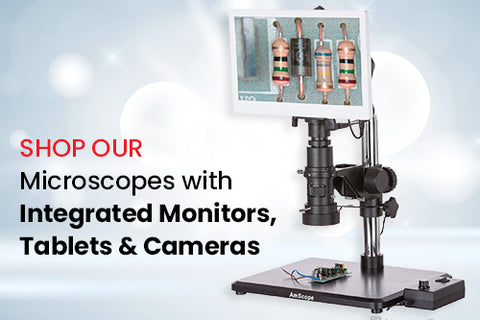
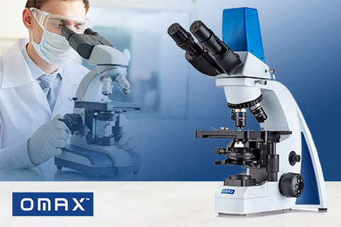
Once you've pinpointed your ideal microscope, you can order one from AmScope. We sell a range of premium light scopes, including student microscopes of various diffraction limits, microscopes for children, and compound microscopes. Besides AmScope products, we also sell scopes from brands like OMAX, IQCrew, and Q-Scope. Additionally, we provide free guides about microscope light bulb maintenance and light microscope parts and functions.
REFERENCES
Boundless. Microbiology.
Leica. "Confocal Microscopes."
MicroscopyU. "Differential Interference Contrast."
MicroscopyU. "Introduction to Phase Contrast Microscopy."
Moyes, Rita B, Reynolds, Jackie, and Breakwell, Donald P. "Preliminary staining of bacteria: simple stains."
News Medical Life Sciences. "What Is Ultraviolet Microscopy?"
Nikon. "Confocal Microscopes."
ONI. "Fluorescence microscopy."
Oxford Reference. "Ultraviolent Microscope."
Wang, Gufeng and Fang, Ning. "Chapter four - Detecting and Tracking Nonfluorescent Nanoparticle Probes in Live Cells."