


How Does a Compound Microscope Work?

Human beings have always been curious about their surroundings. But when people first began to study flora and fauna, their observation of nature was limited to the visible world. Once the concept of magnification was introduced, researchers discovered that there was more than meets the eye.
“Micro” means small and “scope” means to see or view. Thus, humans developed microscopes to observe small things that are invisible to the naked eye. The earliest optical microscopes were only a step up from a magnifying lens. They had simple scopes connected to a single lens. This resulted in limited magnification ability, and they were called simple microscopes.
In 1590, three Dutch spectacle makers invented an optical microscope with more than one lens. This was the first compound microscope. Today, compound microscopes are used in most research laboratories, hospitals, and schools. Read on to learn more about what a compound microscope is and how it works so that you can select the best type of microscope for you.
What Is a Compound Microscope?
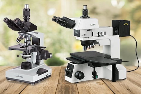
A compound microscope is a type of optical microscope that uses visible light and multiple lenses to help you observe a real and magnified image of tiny objects. Through a compound lens system, it produces enlarged images of microscopic objects like living and dead organisms, tissues, and cells.
Compound microscopes used for studying biological specimens are also called biological microscopes.
The mechanical parts of a compound microscope include:
Foot. A U-shaped base that supports the weight of the microscope.
Arm. A curved projection resting on the base, used for gripping the microscope.
Stage. A flat plate connected to the lower end of the curved arm. It has a hole (aperture) at the center that allows the passage of light. Specimens to be examined are mounted onto the stage.
Clips. These fasten the specimen-containing slides onto the stage to prevent movement during observation.
Diaphragm. Attached below the stage, it can be either a disc or iris diaphragm that controls the intensity of the light entering the microscope.
Pillar. A vertical projection rising from the base that supports the stage.
Inclination joint or pivot point. Connects the arm to the pillar and is useful for tilting the microscope.
Body tube. A hollow tube attached to the upper end of the arm. The upper portion (draw tube) contains the eyepiece lens. The body tube length is usually around 6.3 inches or 160 millimeters.
Nose piece. A metallic disc connected to the base of the body tube that supports the objective lenses.
Adjustment knobs. There are two knobs for fine and coarse adjustment. They move the body tube to bring the object being studied into focus.
Next, let’s discuss the optical parts and functioning of biological microscopes.
How a Biological Microscope Works Using Visible Light
The optical parts of a compound microscope include a mirror, eyepiece lens, and objective lenses.
The mirror is attached to the base of the microscope. It has a plain surface on one side and a concave surface on the other. The mirror reflects rays from an external light source into the microscope.
Compound light microscopes have their own light source (illuminator) attached to the microscope base. The light rays are focused onto the stage by the condenser, which is placed below the stage.
Relies on a Set of Lenses To Increase Optical Resolution and Magnification
Microscope lenses use transmitted, refracted, or reflected light to increase the image size of the object being viewed. This aspect is called magnification. The magnification power of a microscope depends on the lenses’ ability to bend light waves.
The object’s image must not only be enlarged but also its minute details must be clearly visible to the viewer’s eye. This aspect is called resolution. The clarity of the image produced depends on the lens quality and the frequency of the light waves falling onto the specimen. This is because light rays with high frequency have short wavelengths, which improve the image resolution.
Lenses with high magnification power also tend to provide greater resolution. Here are the two main types of lenses in a compound microscope:
Objective Lenses
They’re attached to the nosepiece at the base of the body tube. They can be of many types — scanning (4X magnification), low power (10X magnification), high power (40X magnification), oil immersion (100X magnification), and specialty (2X, 50X oil, 60X, and 100X dry magnification).
Eyepiece Lens
The eyepiece or ocular lens is attached to the top of the body tube. The rim of the eyepiece contains markings such as 5X, 10X, 15X, and 20X, which denote its magnification power. Place your eye above this lens to observe the magnified image of the object placed on the stage.
As Magnification Increases, a More Detailed Image Appears
When rays from an incandescent light source enter the microscope lenses, they’re not uniform. They’re travelling in multiple directions and have differing wavelengths. Once the light waves pass through the specimen and reach the objective lenses, they’re bent into parallel paths. The objective lens then projects an inverted but magnified image onto a fixed intermediate image plane within the microscope. The ocular lens further magnifies this projected image.
Magnification power is calculated as the extent of image enlargement performed by the lens. For example, a magnification power of 10X indicates an approximate 10-fold increase in the size of the object’s image. Because compound microscopes have multiple lenses, the total magnification achieved is a numeric multiple of the magnification powers of all the individual lenses.
Thus, as the image size increases with the magnification power, more details become visible.
Changing the Distance From a Prepared Slide Makes a Difference
Lenses with high magnification power bend light rays much more than lenses with low magnification power. This makes the image of the object come into focus at a much shorter distance from the lens. Hence, lenses with high magnification power are placed much closer to the specimen under observation. Conversely, low-power lenses are placed further away from the prepared slide.
Selecting the Right Type of Microscope for Your Lab
Microscopes have become a critical part of modern science laboratories, whether that may be while you’re still studying in schools and colleges or working at hospitals and research facilities.
With more than 15 years of experience, AmScope offers a variety of best-in-class compound microscopes based on your unique needs.
Through our informative pages, you can also learn more about other types of microscopes like stereo microscopes to see if they’re better suited to your situation.